Techniques

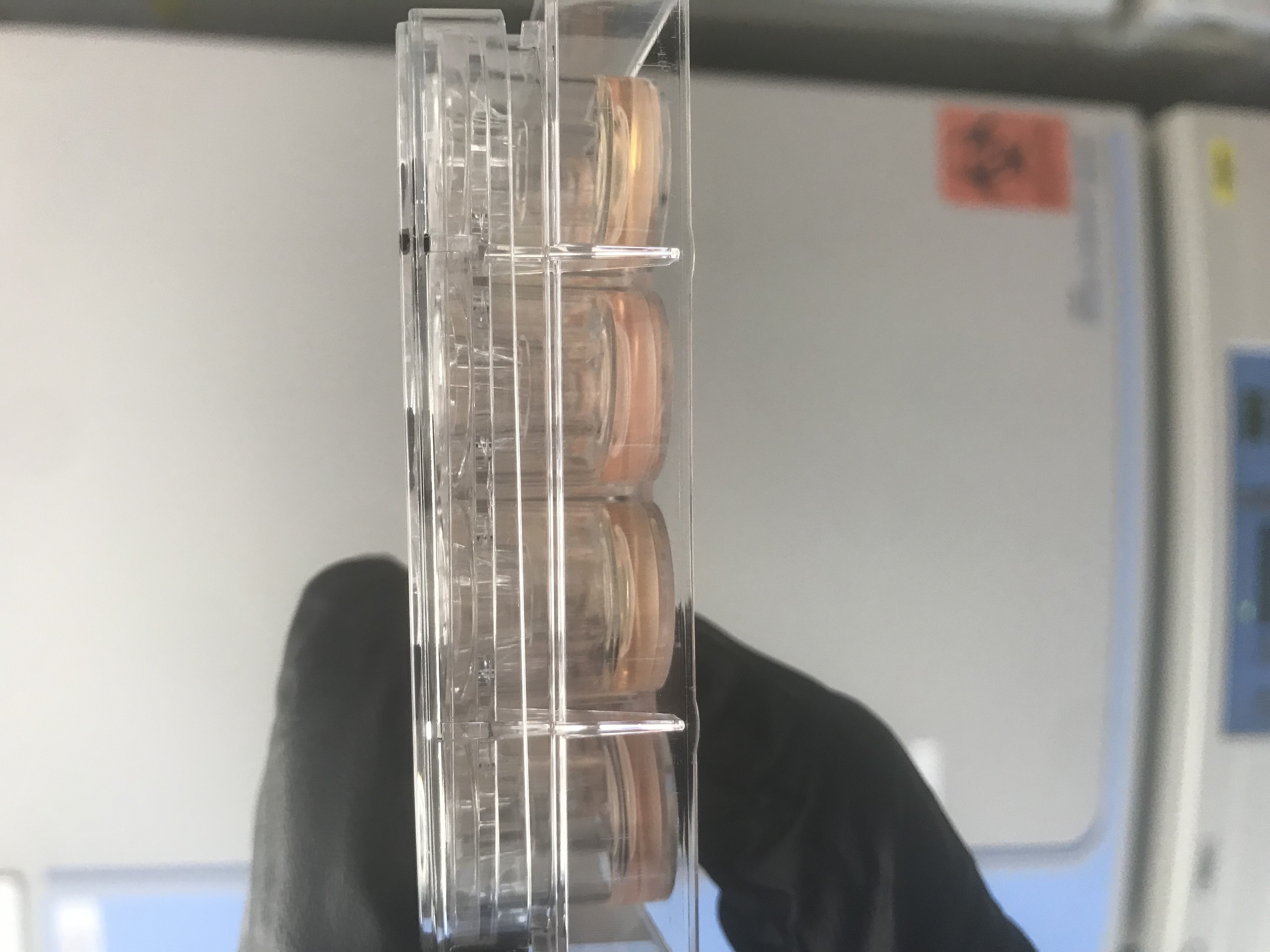
24 Traswell Culture Plate
Ussing Chamber
The Ussing chamber technique, developed by Danish physiologist Hans Ussing in the 1950s, was originally designed to investigate mechanisms of ion transport across frog skin. Ussing integrated electrophysiological tools into the technique, paving the way for precise measurement of transepithelial voltage, current, and conductance. This pioneering method laid the foundation for modern studies of epithelial physiology.
Today, Ussing chambers combined with electrophysiology are widely used to quantify ion transport across epithelial barriers. By mounting tissue or cell monolayers between two chambers and applying a voltage, researchers can measure short-circuit current, which reflects the net movement of ions driven by active transport mechanisms. Alternatively, epithelia can be studied without an applied voltage to model epithelial transport under physiological voltage conditions. In either approach, a voltage or current pulse will provide the transepithelial conductance—a measurement of the ease of ion flow through the epithelium. Overall, the technique enables detailed analysis of transport proteins, ion channels, and the effects of cell signaling, pharmacological agents, or genetic modifications.
For further reading:
Hamilton KL. Ussing's "Little Chamber": 60 Years+ Old and Counting. Front Physiol. 2011
Flow Cytometry
The flow cytometer enables us to determine cell population and gene expression using fluorescently labelled antibodies.
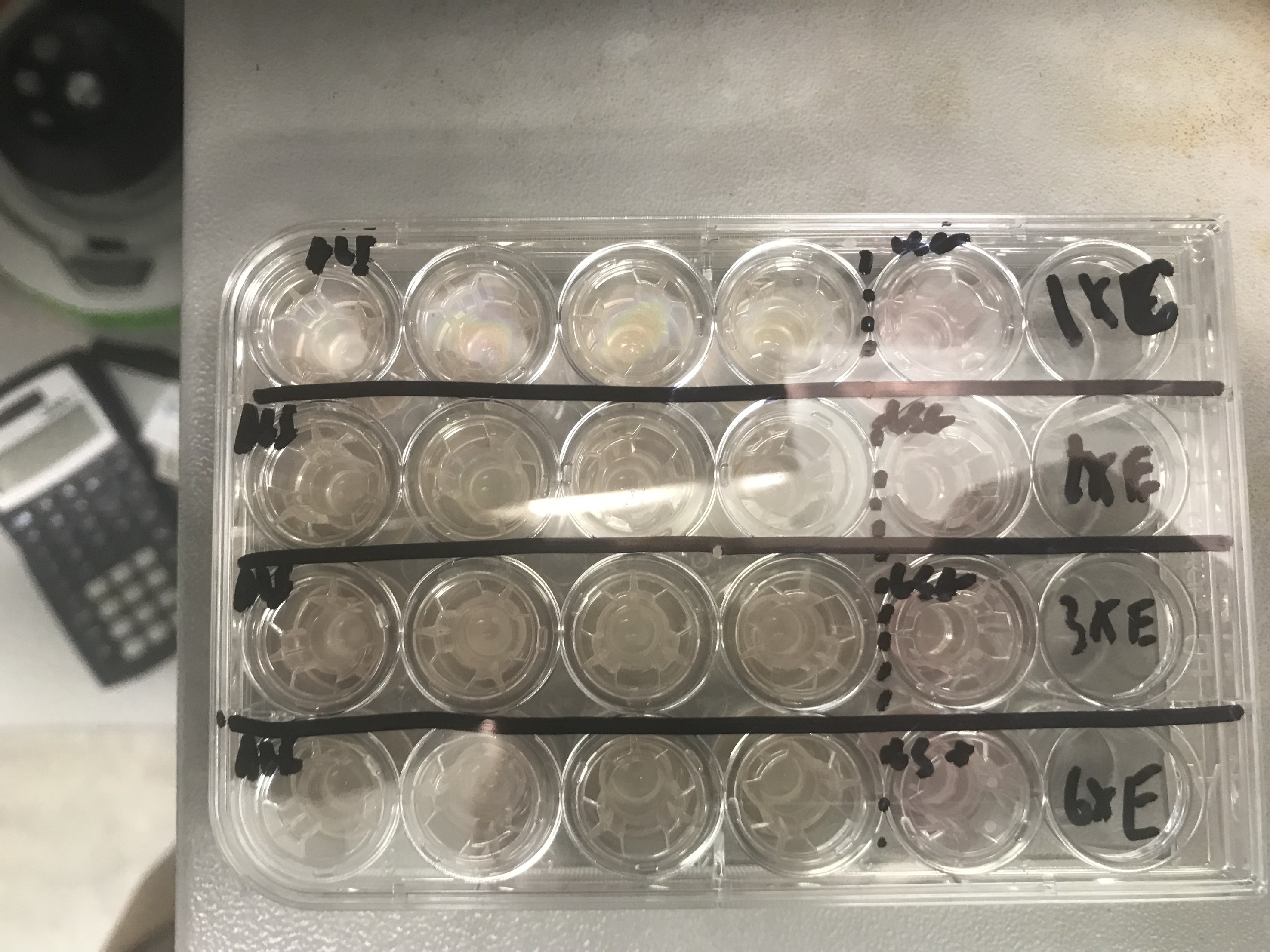
Transwell Culture System
Our transwell culture system enables us to culture differentiated epithelial monolayers which mimic the epithelia present in the airway. These cultures can then be used for Flow Cytometry, Electrophysiology, Immunofluorescence, and many other assays.
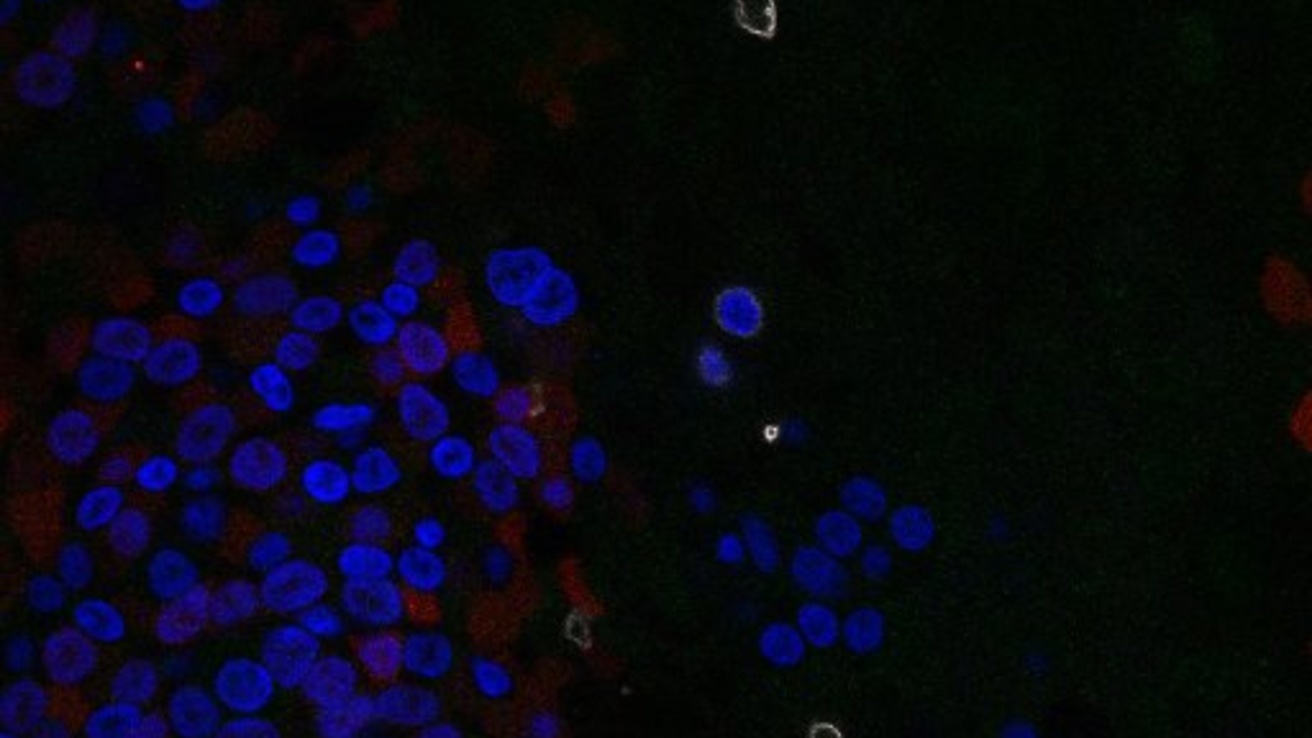
Confocal Microscopy
Our confocal microscope enables us to visualize immunofluorescent labels on our airway cultures. This is invaluable to validate cell/tissue populations and morphology when paired with Flow Cytometry.
Nucleofection
Our nucleofector enables passage of DNA and other molecules into the cell's cytoplasm. We mainly utilize this technology as part of the CRISPR/CAS9 gene modificaiton system.